🔬 Microscope FOV Calculator
Calculate your microscope's Field of View diameter for any objective & eyepiece combination
| Objective | Total Mag (10x eye) | FOV Diameter (mm) | FOV Diameter (μm) | FOV Area (mm²) |
|---|---|---|---|---|
| 1x | 10x | 20.00 | 20,000 | 314.16 |
| 2x | 20x | 10.00 | 10,000 | 78.54 |
| 4x | 40x | 5.00 | 5,000 | 19.63 |
| 5x | 50x | 4.00 | 4,000 | 12.57 |
| 10x | 100x | 2.00 | 2,000 | 3.14 |
| 20x | 200x | 1.00 | 1,000 | 0.785 |
| 40x | 400x | 0.50 | 500 | 0.196 |
| 60x | 600x | 0.33 | 333 | 0.087 |
| 100x | 1000x | 0.20 | 200 | 0.031 |
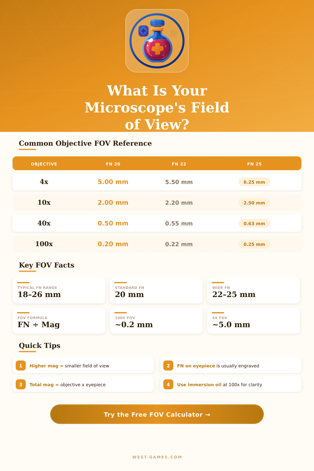
| Objective | FN 18 (mm) | FN 20 (mm) | FN 22 (mm) | FN 25 (mm) | FN 26.5 (mm) |
|---|---|---|---|---|---|
| 4x | 4.50 | 5.00 | 5.50 | 6.25 | 6.63 |
| 10x | 1.80 | 2.00 | 2.20 | 2.50 | 2.65 |
| 20x | 0.90 | 1.00 | 1.10 | 1.25 | 1.33 |
| 40x | 0.45 | 0.50 | 0.55 | 0.63 | 0.66 |
| 60x | 0.30 | 0.33 | 0.37 | 0.42 | 0.44 |
| 100x | 0.18 | 0.20 | 0.22 | 0.25 | 0.27 |
| Camera Sensor | Sensor Width (mm) | FOV @ 10x (mm) | FOV @ 40x (mm) | FOV @ 100x (μm) |
|---|---|---|---|---|
| 1/4" CMOS | 3.60 | 0.360 | 0.090 | 36 |
| 1/3" CMOS | 4.80 | 0.480 | 0.120 | 48 |
| 1/2.5" CMOS | 5.76 | 0.576 | 0.144 | 57.6 |
| 1/2" CMOS | 6.40 | 0.640 | 0.160 | 64 |
| 1/1.8" CMOS | 7.18 | 0.718 | 0.180 | 71.8 |
| 2/3" CCD | 8.80 | 0.880 | 0.220 | 88 |
| 1" CMOS | 13.20 | 1.320 | 0.330 | 132 |
| Micro 4/3 | 17.30 | 1.730 | 0.433 | 173 |
Note: This text bases on real descriptions and precise technical details about the FOV of microscope.
The FOV in microscope, usually called FOV show the part of the sample, that one can observe during one moment through the device. It forms a round area, visible through the eyepiece. In short words, FOV estimates the size of the image, that the observer notices.
What the Microscope Field of View Is and How to Measure It
Usually one points it in millimeters, and it is useful for estimate the size of the object, that one studies.
Optics of the microscope delivers clear and sharp sight around the optical axis. From that zone one chooses the FOV. If the FOV is below one millimeter, one commonly uses micrometers, or also microns, for the reading.
One millimeter matches to thousand units of microns.
The number of the FOV, sometimes marked as F.N., points the diameter of the viewing area in millimeters, measured in the central plane of the image. It determines the scope of the sample, seen through the opening of the eyepiece. Diameter of the FOV depends on the magnification grade of the objective.
During usage of the combination eyepiece-objective, the FOV of the objective expands because of the eyepiece.
For estimate the FOV, the process is fairly easy. One divides the number of the FOV by the magnification reading. For instance, if the eyepiece shows 10x/18 and the objective has 40-times magnification, first multiply 10 by 40, too receive 400.
Later divide 18 by 400, what gives diameter of 0.045 millimeters for the FOV. Additional sample: dividing the number 20 by a 10x-objective, one gets 2 mm of diameter for the FOV.
Usually the weakest magnification delivers the most wide FOV. However the kind of eyepiece also matters. There are wide-field and even broader variants, that intend to offer bigger areas for viewing.
Some costly microscopes use eyepieces with 30 mm diameter, so the field can extend more.
Here some useful details about cameras and FOV. The FOV of microscope forms a circle, while that of a camera is rectangular form, that must fit in the circle. Even at ideal fitting, where the points of the rectangle touch the edges of the circle, only around 50 percent of the circular area shows.
More small sensors of cameras make the situation even harder. One can estimate the FOV for digital systems, dividing the diagonal size of the sensor by the magnification of the objective lens.
For well understanding reasons about FOV during choice of a fitting microscope. If one knows the FOV, easily estimates one outside, how much sample shows with a particular objective. Marks on eyepieces as WF10 or WF25 point the magnification.
Calibration slides with little scales onthem allow to measure the real FOV directly.